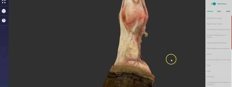
Equine Distal Forelimb Dissection in 3D: Nerves and Vessels
A scan of a real dissected equine distal forelimb, with remarkable detail of the nerves, arterial supply and venous drainage of the limb.

Ivalalearn
Proud to be part of the Veterinary Information Network family

A scan of a real dissected equine distal forelimb, with remarkable detail of the nerves, arterial supply and venous drainage of the limb.
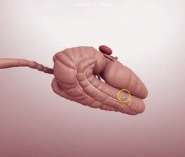
Our equine abdomen content focuses on the gastrointestinal tract, so you can quickly understand the caecum and the different parts of the colon.
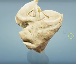
A scan of the proximal tibia of a cow, which our anatomists say is extremely similar to the horse, to help you understand the ligaments of the stifle.
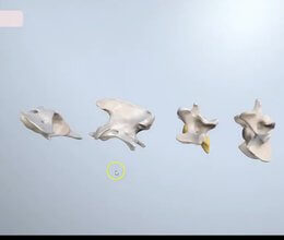
We refreshed our canine spine with higher quality scans and whole-region landmarks, and added an individual thoracic and lumbar vertebra to explore.
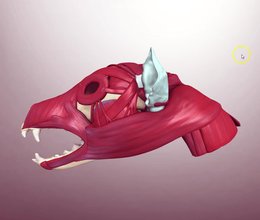
Our canine skull musculature content covers the origins and insertions of the head muscles, lets you sort them by nerve, and has Test Yourself built in.
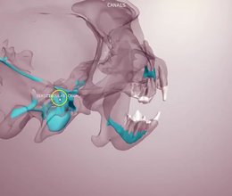
Our canine skull content in four scenes: bony landmarks, teeth and foramina, the canals inside a transparent skull, and a schematic of the cranial nerves.
Ivalalearn
Proud to be part of the Veterinary Information Network family
OUR MISSION:
To shape more confident students and clinicians by creating intuitive, engaging, interactive learning content.
OUR MISSION:
To shape more confident students and clinicians by creating intuitive, engaging, interactive learning content.
Ivala is proud to be a wholly owned subsidiary of the Veterinary Information Network (VIN)
IVALA ® All rights reserved.



